shoulder pain breast cancer
 Breast Cancer Awareness: Shoulder Pain - Jaspal Ricky Singh, M.D
Warning: The NCBI website requires JavaScript to operate. The shoulder pain due to metastatic breast cancer -A young report-salir case KimDepartment of Anesthesiology and Pain Medicine, Faculty of Medicine, Keimyung University, Daegu, Korea. Min Woo JungDepartment of Anesthesiology and Pain Medicine, Faculty of Medicine, Keimyung University, Daegu, Korea. Jin Mo KimDepartment of Anesthesiology and Pain Medicine, Faculty of Medicine, Keimyung University, Daegu, Korea. Abstract A broken rotating cuff causes shoulder pain and limits the movement of the shoulder joint. A chronic degenerative change or impingement is the reason for a broken rotating cuff. The diagnosis is made based on medical history and physical and radiological examinations. Other causes of shoulder pain include calcific tendonitis, degenerative arthropathy, joint dislocation, primary or metastatic fracture and neoplasm. However, metastatic cancer in the shoulder joint is difficult to diagnose. We experienced a case in which a 46-year-old patient complained of left shoulder pain and limited joint mobility, and these symptoms were due to metastatic breast cancer on the shoulder. The shoulder pain occurs in 6.6 to 25 people of every 1000, and is the third most common musculoskeletal disease after low back pain and knee pain [-]. The shoulder joint is the joint with the highest range of movement in the body, and the cause of shoulder pain is not only limited to the shoulder joint, but can also be caused by injuries in the surrounding area. Common causes of shoulder pain are rotating fist pathology, adhesive capsulitis, calcific tendinitis, degenerative joint disease, dislocation, fracture, acute trauma and tumors []. The authors have recently experienced a case in which a 46-year-old patient complained of left shoulder pain and movement limitation (LOM). After taking a medical history and giving a physical exam, it was suspected that the pathology of the rotating cuff with tear of the rotator cuff, but no anomalies were observed in ultrasound. Through magnetic resonance imaging (MRI) and bone scan, metastatic breast cancer that was believed to have been completely removed by prior surgery was observed in the wet head. In this case, we report a case of metastatic breast cancer in the shoulder joint's humeral head in a 46-year-old woman along with a literature review. CASE REPORT The 46-year-old patient visited our department that complained of left shoulder pain that began after the frequent use of arm 1 month before his visit. The pain was dull in nature with a visual analogue scale (VAS) of 35/100 mm and when the left arm was moved, the pain was increased with a 100/100 mm VAS. He also complained of the LOM in the left shoulder joint and pain sleep interruption. The patient had been diagnosed with right breast cancer 6 years before his visit and had made a modified radical mastectomy. He had been regularly visiting a surgical clinic until recently, was taking tamoxifen every day, had a bone scan and body ultrasound done in the breast area once a year, and PET-CT (Positron Emission Tomography-Computed Tomography) took once every two years to observe progress, but there was no trace of recurrence or metastasis. In the physical examination, there were no irregularities in the inspection, but there was tenderness in the left palpado greater tuberosity. The LOM in its left shoulder joint during the active movement was the following: 150° bending and 50° extension of the sagittal plane; 110° abduction, 50° aggregation of the coronal plane; 40° external rotation. As for internal rotation on the back, your left hand could reach your first lumbar vertebrae. Meanwhile, in terms of passive movement, the patient complained of pain between 90 to 120° in kidnapping, but there was no LOM in any direction. The patient was positive for the Neer test, positive for the empty tin test, and positive for the test of the gout arm; therefore, the pathology of the rotating cuff was suspected with a rupture of the accompanied supraspinatus tendon. Therefore, a laboratory and simple x-ray examination was performed, but there were no anomalies, and the left shoulder area was examined using ultrasound by our department, but the pathology of the anticipated rotating fists could not be confirmed. An ultrasound-guided left-handed suprascapular block was performed to treat pain in the shoulder area, but pain relief was insignificant. Therefore, the Department of Radiology was asked to do an echosonography, but no abnormalities were found in the rotary cuff. The patient complained of continual pain and LOM to make a magnetic resonance and bone scan. The MRI examination saw a cancerous lesion in the humeral head (), and the bone scan found suspicious abnormalities of a metastatic tumor in the humeral head and the 10th chest vertebrae. The patient was transferred to the Department of Internal Medicine and was diagnosed with metastatic breast cancer in the wet head and 10 chest vertebrae and received chemotherapy and radiation therapy. Currently, six months later, there are no observations of metastases in other areas and the cancer lesion in the head of the wetland has not changed. Magnetic resonance imaging. The axial image weighted by T2 (A) and the coronal image (B) shows osteolytic lesion in the left head and humeral metaphysics. DISCUSSION The rotary cuff is composed of the supraspinatus, infraspinatus, subscapularis and minor teres. These ensure the shoulder joint so that it can move and allows the normal functioning of the shoulder. However, the rotary cuff is worn easily and susceptible to degeneration, so it is the weakest part of the shoulder joint. Therefore, tears of the rotating cuff occur frequently, and this weakens the shoulder joint and causes pain []. A rotating cuff tear occurs from the mixed effects of various mechanisms such as mechanical impact, degenerative changes, circulatory disorders and joint abrasion. Miller and Dlabach [] contend that the supraspinatus is more susceptible to tear in the rotating cuff. The tear of the supraspinatus tendon occurs from the abrasion by being trapped between the humeral head and the acromion or acromioclavicular joint instead of mechanical impact [,]. For the diagnosis of tear of the rotating cuff, history taking and physical examination are important. The patient finds it difficult to lift the arm and is characteristic that the active movement is limited but the passive movement is free in the physical examination. Also, when the arm is lowered, the sign of the drop arm appears where the patient does not have the strength or the arm drops due to the pain. The findings of a physical examination are different for the tears of each tendon. In a supraspinatus tear, a painful arch sign is detected between 60 and 120° in the abduction. When the arm rises to a certain level, the final lift can be performed easily. Also, when both arms extend around 40° and the elbow joints extend to where the thumbs point down as if pouring water out of a cup, the pain is generated when the resistance applies to the upper area (force can prove). In an infraspinatus tendon tear, when the arm rotates passively to the maximum range of external rotation, the patient is not able to maintain this active position and the arm is removed from the body or rotated internally (load sign). In a subscapular tendon tear, when the patient's arm is behind the back, there is a lack of muscle strength that makes it difficult to stretch from the back as it overcomes the resistance (extract the test) []. The radiological diagnostic method for tears of rotary cuff uses ultrasonography or magnetic resonance examination. Rafii et al. [] reported that the sensitivity of tears of rotating cuff in the MR was 95% in the case of tears of full thickness and 84% in the case of partial tears, while Quinn et al. [] reported that the sensitivity of magnetic resonance examinations in relation to tears of rotating cuffs was 84% and the specificity was 97%. Burk et al. [] compared the MRI observations and arthrography with direct observations during surgery in 16 cases and reported that MRI and arthrography had sensitivity of 92% and 100% specificity. There are also reports on the usefulness of ultrasound in tears of rotating cuff, in which Brandt et al. [] reported that, compared to observations during arthroscopic surgery in 38 cases, ultrasound had 57% sensitivity and 76% specificity. As you can see, ultrasound is known to be less accurate compared to magnetic resonance in the diagnosis of tears of rotating cuff. Ultrasound has many merits in which it is possible to perform tests and treatment in real time; there is no risk of radiation exposure so that it can be safely used in pregnant women and children, and it is cheap and non-invasive. However, there are limitations to ultrasound such as seeing deep structures such as intestines because it cannot penetrate the air and cannot pass through the bone or metal, so there are limits to evaluating the bone marrow or the safety of the orthopedic hardware. The most common malignant neoplasm in women is breast cancer, taking 32% of malignant neoplasms. With the development of modern diagnostic techniques and treatment modalities, the mortality rate is decreasing, but it remains the most common cause of death for women in their forty years. Coleman and Rubens [] claim that the complete recovery of breast cancer is possible if surgically removed at the initial stage, and that bone metastasis occurs in 69% of all cases of breast cancer. According to Martin and Moseley [], breast cancer is easily spread to the spine, rib, pelvis and proximal long bone, and the metastasis rate is 70%. Thomas et al. [] reported that cancer cells that spread to the bone marrow accelerate osteolysis and osteoclast reactions and may cause pathological fractures or pain. In the shoulder pain caused by metastatic cancer, cases of lung cancer are reported that has spread to humerus [], and kidney cancer that has spread to humerus and escapula []. Computed tomography, magnetic resonance and bone scan are used to diagnose cancer that has spread to the bone, and the irregular osteolytic lesions that appear in these images are the most common observations for skeletal metastases []. The patient in our study had received surgery in the past for breast cancer, but had not shown trace of recurrence or metastasis in observations of progress through regular visits. Since he complained of pain on the left shoulder, he had limited active joint motion, but free passive joint movement, and was positive for the sign of the gout arm and the vacuum can prove, pathology of the rotating cuff with a tear of the supraspinatus tendon was suspected; however, there were no images indicating the tear in ultrasonography images. Thus, magnetic resonance and bone scan were performed and, as a result, breast cancer was found that spread to the bone. Recently, the use of ultrasound in pain clinics increases. However, it is necessary for health professionals to realize that ultrasound cannot penetrate the bone and that additional tests such as magnetic resonance may be needed when there is discordium with clinical symptoms. In addition, cancerous lesions or skeletal metastases should be considered when musculoskeletal pain occurs in patients who think they have recovered completely from the cancer. ReferencesFormats: Share , 8600 Rockville Pike, Bethesda MD, 20894 USA

Shoulder Pain and Breast Cancer - CONQUER: the patient voice

Side effects of radiation for breast cancer: What to know
Shoulder Pain after Breast Cancer Treatment | Bangalore Shoulder Institute

Shoulder Pain after Breast Cancer | Physio Body and Sole

Breast Cancer Symptoms and Early Warning Signs

Lymphoedema: causes, symptoms and treatment | Breast Cancer Care

12 Effects of Breast Cancer on the Body

Evaluation and Management of Breast Pain: Dennis R. Holmes, M.D., F.A.C.S.: Breast Cancer Surgeon

Pin on Health

5 signs of breast cancer that aren't a lump | CTCA

Neck, Shoulder and Arm Pain following Breast Cancer Treatment | Active Life Physiotherapy Carlow

Breast pain: Causes, types and treatments | Breast Cancer Now
Shoulder pain in a patient with cancer — NUEM Blog
/shoulder-pain-56a82a0b3df78cf7729cb497.jpg)
Shoulder Blade Pain: Symptoms, Causes, Diagnosis, and Treatment

Early Signs of Breast Cancer, From 9 Women With the Disease | Health.com
![PDF] The Shoulder Pain due to Metastatic Breast Cancer -A Case Report- | Semantic Scholar PDF] The Shoulder Pain due to Metastatic Breast Cancer -A Case Report- | Semantic Scholar](https://d3i71xaburhd42.cloudfront.net/4bd132ef912ad4c594b6ba24371acc7ba3bb3037/2-Figure1-1.png)
PDF] The Shoulder Pain due to Metastatic Breast Cancer -A Case Report- | Semantic Scholar
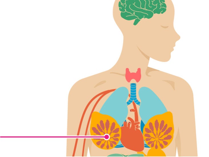
12 Effects of Breast Cancer on the Body

Bone cancer: Survival rate, causes, types, and treatment

Breast Cancer and Pain: Why Treatment Can Cause Arm and Shoulder Pain

Breast Cancer and Shoulder Blade Pain: What You Should Know

Breast Cancer (6): Researching My Options (Leonie Saysell) | leoniesaysell
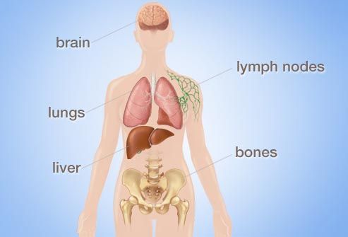
Where Breast Cancer Spreads: Lymph Nodes, Bones, Liver, Lungs, Brain

Shoulder Pain: 12 Reasons Your Shoulder Hurts | Health.com
:max_bytes(150000):strip_icc()/causes-of-left-breast-pain-429842___-02-5b4e6ac346e0fb005b240a36.png)
What's Causing My Left Breast Pain?

Shoulder Blade Pain - Possible Causes and Home Treatments

Breast Cancer vs. Cyst: Symptoms, Causes, Treatment & Prognosis

Breast Cancer and Pain: Why Treatment Can Cause Arm and Shoulder Pain

Are there other signs of breast cancer? | Norton Healthcare Louisville, Ky.

Ouch! Shoulder pain and how to treat it - Harvard Health

Shoulder blade pain: Causes and treatment

Surprising Lung Cancer Symptoms

Joint Pain: A Common Side Effect of Certain Breast Cancer Treatments – Health Essentials from Cleveland Clinic

Kathy: Neck Pain and Breast Cancer - Feldenkrais Method in Kirkland and Seattle

Representative image of shoulder ultrasonography in patients with... | Download Scientific Diagram

12 Effects of Breast Cancer on the Body
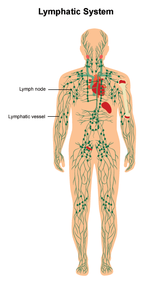
Lymphedema | Cancer.Net

How to release a frozen shoulder - Harvard Health

Why Does My Shoulder Hurt? - OrthoRehab - Edina Physical Therapists
/GettyImages-758309241-5b25ab92303713003617a52e.jpg)
Causes of Itchy Breasts Beyond Breast Cancer

Breast Cancer & Delayed Radiation Injury: Symptoms & Solutions
Posting Komentar untuk "shoulder pain breast cancer"